Figure 9.
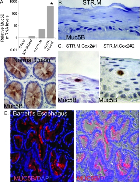
Intestinal and subepithelial gland mucin Muc5B induced by Cox2 expression in STR cells. (A) Quantitative SYBR Green RT-PCR analysis of Muc5B mRNA expression in STR.M or STR.M.Cox2 cells cultured under normal two-dimensional conditions or under three-dimensional organotypic conditions (OTSTR.M and OTSTR.M.Cox2). ΔCt values and statistical analysis were calculated as before after duplicate PCRs for each sample, n = 4. *Significantly differs from all other cells, P < .01. NS indicates nonsignificant difference with STR.M or STR.M.Cox2. (B) Negative immunohistochemical staining for Muc5B in STR.M control cells in organotypic cultures. (C) Muc5B immunohistochemical staining localized to the mucin-containing cysts in organotypic cultures of both STR.M.Cox2 cell lines (#1 and #2). (D) Pattern for Muc5B expression in normal human colon by immunohistochemical staining. (E) Epifluorescent staining for Muc5B (red) in human Barrett epithelium biopsy section. Nuclei counterstained with DAPI (blue). Shown on the right is a merged image of the fluorescent channels with a differential interference contrast image of the tissue.

