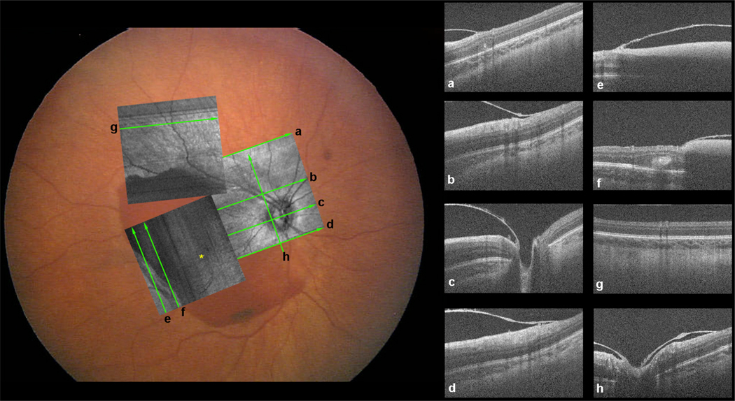
Figure 1.
RetCam photo with spectral domain optical coherence tomography (SD OCT) fundus image overlaid on top after image registration. The lines show the locations of the SD OCT scans in the boxes to the right; each line and SD OCT image has a corresponding label. The OCT images show the retinoschisis cavity with sub-internal limiting membrance (ILM) hemorrhage extending to the optic nerve.

