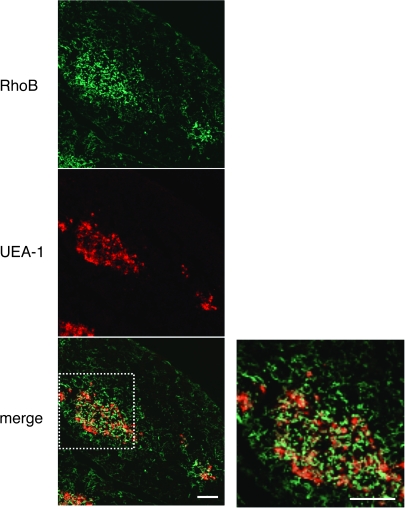
Fig. 1.
Expression of RhoB in mouse thymus. Immunofluorescence staining of thymus sections of 6-week-old C57BL/6 mice was performed to detect RhoB (green) and the binding to thymic medullary epithelium marker UEA-1 (red). Right panel: high magnification from white box in the left panel. Double labeling for RhoB and UAE-1 shows their co-localization, indicating that thymic medullary epithelium expresses RhoB. Data are representative of at least three independent experiments with more than five mice. Scale bars = 100 μm.

