
Fig. 1.
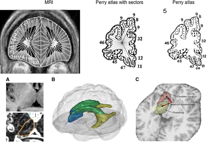
Anatomical methods: stereotaxic and Manual tracings. Upper panel Left Coronal MRI showing application of 20 lateral and 10 medial sectors. Center and Right Perry atlas with same sector algorithm applied (see “Methods”). Lower Panel a Axial view of an MRI slice before (above) and after (below) applying the Sobel filter. This method allows distinguishing the boundaries of the different subcortical structures, especially the thalamus, whose tracing is shown. Yellow arrow indicates lateral edge of thalamus and blue arrow enhanced anterior horn of ventricle. b Three-dimensional model merged from the manual tracings of the lateral ventricles. The anterior horn is depicted in blue (note blue arrow indicating edge in a), the lateral horn is in green, and the temporal horn appears in yellow. c Location of the centers of mass of the caudate nucleus and thalamus, and vectors showing their position regarding to the anterior commissure in the midline of the brain (yellow arrow for orientation to a)