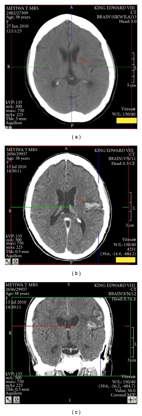
Figure 2.

(a) Axial computerised tomography (unenhanced) brain showing a wedge-shaped hypodensity in the left frontopareital area with extension across both gray and white matter, which is in keeping with a cerebral infarct. (b) Axial computerised tomography (enhanced) brain showing hyperdensity in the left-hand side in the area of previous ischaemia. (c) Coronal computerised tomography (enhanced) brain showing hyperdensity in the left-hand side in the area of previous ischaemia.
