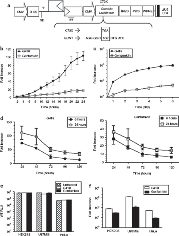
Figure 2.
Regulation of Gaussia luciferase expressions in cells transduced by HIV-2 based lentiviral vectors. (a) Schematic representation of the sequence and location of the inserted premature termination codon (PTC) within the Gaussia luciferase (hGLuc) gene along with the Tat-independent, self-inactivating HIV-2 based lentiviral vector used. ψ, packaging signal; SD, major splice donor; CMV, cytomegalovirus immediate early gene promoter; puro, puromycin resistance gene; WPRE, woodchuck hepatitis virus post-transcriptional regulatory element; rre, rev response element; LTR, long-terminal repeat. (b,c) Kinetics of turning “On” luciferase expression in human embryonic kidney 293 (HEK293) cells infected with the HIV-2 lentiviral vector harboring the mutant hGLuc gene. 1 × 10⁴ stable mass population of transduced HEK293 cells were seeded in 96-well plates and exposed to a constant concentration of 400 µg/ml of G418 (circles) or 1 mg/ml of gentamicin (squares). Luciferase expression was measured from the cultured media every 2 hours up to 24 hours and then every day up to 6 days. (d) Kinetics of turning “Off” luciferase expression in HEK293 cells transduced with lentiviral vector harboring the mutant hGLuc gene. 2.5 × 10⁵ stably selected mass population of transduced cells were seeded in 6-wells and exposed to a constant concentration of 400 µg/ml of G418 (circles) or 1 mg/ml of gentamicin (squares) for 6 hours or 24 hours. Cells were washed with phosphate buffered saline at time of aminoglycosides removal and fresh media was added to the cell. Cultured media was assayed for luciferase activity at 24-hour intervals. (e,f) Luciferase expression in various cell lines transduced with the HIV-2 lentiviral vector harboring the wild-type or mutant allele of hGLuc. Infected cells were selected for puromycin resistance to obtain a stably transduced mass population of cells. Then 2.5 × 10⁵ cells were exposed to 150 µg/ml of G418 and 1 mg/ml of gentamicin for 48 hours before luciferase expression was measured. Wild-type luciferase activity was measured in relative light units (RLU). Induction of mutant hGLuc was represented as fold induction relative to untreated cells. Luciferase readings were normalized to total protein. All experiments were done in triplicate and repeated at least twice. Error bars represent the standard deviation.

