
Figure 1. FAK deletion impairs recruitment of cardiac neural crest-derived SMC to the developing aorta and pulmonary artery.
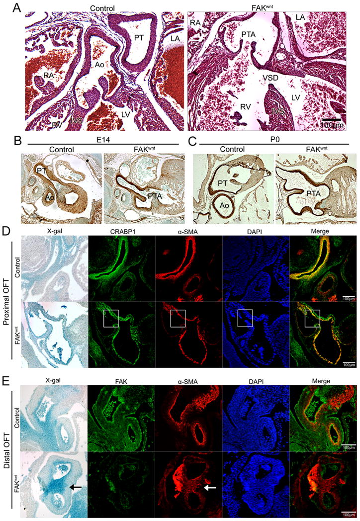
(A) H&E staining of genetic control and FAKwnt mice at postnatal day 0 (P0). FAKwnt outflow tract (OFT) shows persistent truncus arteriosus (PTA) and ventricular septal defect (VSD). LA, left atrium; RA, right atrium; Ao, aorta; PT, pulmonary trunk; IVS, intraventricular septum. (B-C) SM-22 immunostaining (brown) of embryonic day 14 (E14; B) or P0 (C) genetic control and FAKwnt OFT revealed PTA without defect in SMC differentiation. (D) Proximal OFT of genetic control and FAKwnt hearts at E 12 reveals co-localization of α-SMA (red) with the CNC lineage marker CRABP1 (green), indicating that the SMC covering the aorta and pulmonary artery were derived from CNC cells. X-gal staining demarcates wnt-1-derived CNC in the Rosa26RLacZ –positive embryos. Nuclei (blue) were stained with DAPI. Note the lack of SMC coverage (highlighted in white boxes) in the inner walls of the aorta and pulmonary artery in FAKwnt mice. (E) Distal OFT at E 12 stained with X-gal (blue in bright field), FAK (green), and α-SMA (red) reveals FAK-depleted CNC differentiate into SMC (arrows) but fail to appropriately populate the conus. Nuclei (blue) were stained with DAPI. Scale bar = 100μm.