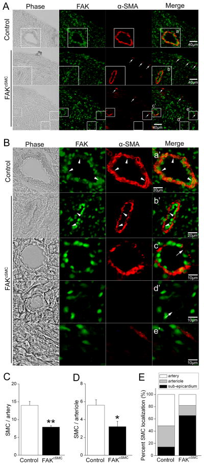
Figure 2. FAK deletion impairs recruitment of epicardial-derived SMC to the coronary vessels.
(A) P0 genetic control and FAKcSMC heart stained with FAK (green) and α-SMA (red). Phase image is shown on left. Sub-epicardial α-SMA-stained cells (arrows) are present in FAKcSMC but not genetic control hearts. Scale bar = 40μm. Boxed regions (a'-e') in A are shown at higher magnification in B. (B) FAKcSMC hearts exhibited reduced presence of SMC lining the coronary vasculature, including large vessels (arrowheads) and small arterioles (arrows), in comparison to genetic control vessels. FAK deletion was confirmed by absence of FAK staining in SMC from FAKcSMC hearts (arrowheads). Scale bar = 20μm or 10μm. (C-E) Numbers of SMC lining arteries (diameter > 65μm), arterioles (diameter < 65μm as previously described45), or localized in the sub-epicardium were quantified using Image J. *p < .05; **p< .01.

