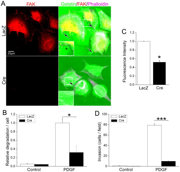
Figure 6. FAK is necessary for SMC-mediated extracellular matrix degradation.
(A) LacZ- or Cre-infected fakflox/flox SMC were plated on Oregon Green 488 gelatin/FN matrix in serum-free medium and treated with vehicle or PDGF-BB for 90 minutes. Cells were fixed and co-stained with anti-FAK antibody and phalloidin. (B) Cells were scored for the presence of degradation puncta (black spots) and data are presented as puncta/per cell normalized to values for the LacZ-infected SMC following PDGF treatment. Data represent mean±SEM of at least 200 cells from 3 independent experiments. *p< .05. (C). LacZ- or Cre-infected fakflox/flox SMC were plated on DQ-gelatin-coated 96-well plate and treated with PDGF-BB for 90 minutes. Fluorescence intensity was monitored at Ex/Em 495/515 nm. Data represent mean±SEM of 3 independent experiments. *p< .05. (D) GFP and LacZ or Cre co-infected fakflox/flox SMC were Cells treated as above were plated on matrigel-coated inserts (10 μg/ml; Bio-Coat) in serum-free media using either PDGF-BB (20 ng/ml) as the chemoattractant. Invading cells were counted by direct fluorescence at 10X magnification. Data represent mean±SEM of 3 independent experiments. ***p< .001.

