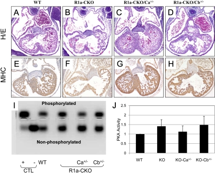
Fig. 2.
Analysis of embryonic hearts at e11.5. A–D, Midlevel sections from WT, R1a-CKO, R1a-CKO/Ca+/−, and R1a-CKO/Cb+/− hearts stained with hematoxylin and eosin (H/E). E–H, Sections immunostained for myosin heavy chain (MHC). Note that hearts are all oriented the same, with the LV in the bottom right corner of each photomicrograph. I and J, PKA assay from control samples and e11.5 hearts of the indicated genotypes. I, Transilluminator photograph of representative PKA assay showing the resolution of phosphorylated and nonphosphorylated PKA substrate. Control (CTL) lanes show PKA-C incubated in vitro with (+) or without (−) exogenous cAMP in the assay. J, Quantitation of the phosphorylated substrate bands from three independent assays. Each assay was normalized to WT PKA activity, which was assigned a value of 1.

