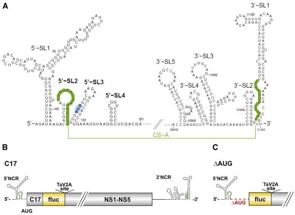
Fig. 1.
Genome secondary structure and organization of TBEV and derived replicon constructs. (A) The 5′ and 3′ regions of the TBEV genome. The stem-loop structures are shown as predicted for the linear form, without consideration of the long-range interaction of the cyclization sequences (CS) (depicted in green) in the viral5′-SL2 and 3′-SL1. The CS element was characterized in an earlier study (Kofler et al., 2006) and was originally called CS-A. The viral start codon AUG within the 5′-SL3 structure is highlighted in blue. (B) Schematic diagram of the parental replicon C17 (wt), in which the structural protein region of TBEV was replaced by an in-frame insertion of the firefly luciferase gene (fluc) (Rouha et al., 2010). C17, truncated capsid protein gene; NS1–NS5, the coding region for the non-structural proteins 1–5; NCR, non-coding region; TaV2A site, Thosea asigna virus 2A site. (C) Schematic diagram of mutant replicon ΔAUG, which has a deletion starting from nucleotide position 133, causing it to lack the entire region encoding the capsid protein, the 5′-SL3 element, and the viral start codon. Diagrams A–C are not drawn to scale.

