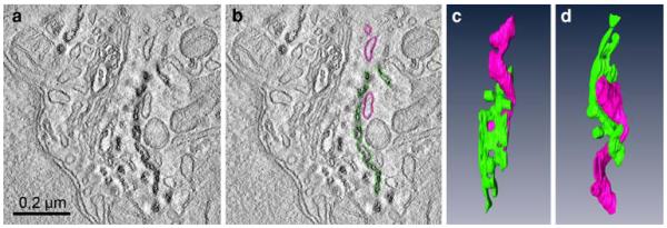
Fig. 9.
Three-dimensional reconstruction of a trans-Golgi-ER junction in a HeLa cell labelled with BODIPY-cer at 4°C for 30 min, followed by postincubation at 37° for 10 min. a and b shows a virtual 2 nm thick slice with (a) and without (b) coloured contours: c and d present the respective model in two different aspects. The trans-Golgi cisterna, defined by reaction product is coloured green, the negative ER cisterna in pink. The different aspects of the 3D-model (c, d) provide insight into the complex helical organization of the junction. Tubular parts of the trans-Golgi ER wind around the multiply perforated trans-Golgi cisterna

