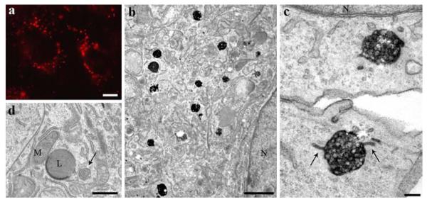
Fig. 1.
Correlative fluorescence and electron microscopic localization of HDL. HepG2 cells were incubated with 50 μg/ml HDL-Alexa568 for 3 h. In the fluorescence microscope, HDL is found in bright, large endocytic compartments enriched in the perinuclear area (a). After DAB-photooxidation, numerous MVBs are positive for internalized HDL (b). Their matrix and the intraluminal vesicle membrane are heavily stained (c). Tubular appendices are frequently positive (arrows). Control cells incubated without labeled HDL were equally processed according to the photooxidation protocol. Cellular compartments including MVBs (arrow), mitochondria (M), and lysosomes (L) are devoid of DAB precipitate (d). Primary magnifications: 1,000× (a), 8,000x (b), 90,000x (c), 10,000x (d). Bars 5 μm (a), 1 μm (b), 100 nm (c), 500 nm (d); N nucleus

