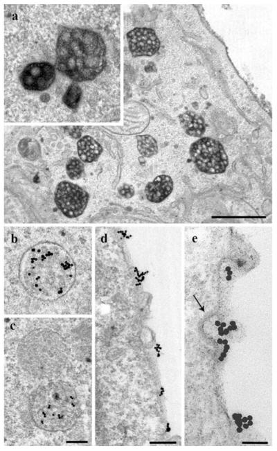
Fig. 2.
Localization of HDL-HRP and HDL-gold. After 3-h internalization, HDL-HRP marks a multitude of MVBs (a). Homotypic fusions of stained MVBs different in sizes and shapes are present (inset). Incubation with HDL-gold for 3 h shows a comparable staining of endosomal compartments and enrichment in MVBs (b, c). HDL-gold positive and negative MVBs may be found side by side (c). With the HDL-gold system discrete PM domains (d, e) and binding to clathrin-coated pits are discerned (arrow; e). Primary magnifications: 17,000x (a), 28,000x (b, c), 40,000x (d), 80,000x (e). Bars 1 μm (a), 250 nm (b, c), 200 nm (d), 100 nm (e)

