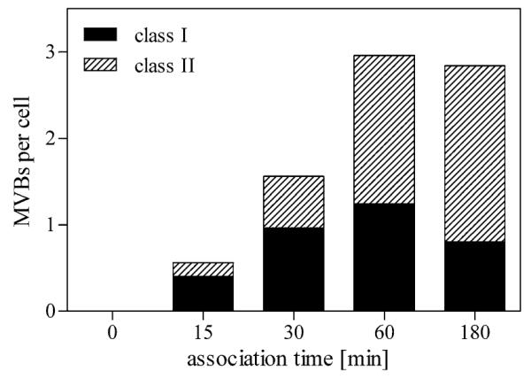
Fig. 4.

Semi-quantitative analysis of HDL-positive MVBs during a 3-h time course. The number of stained MVBs after HDL internalization and DAB-photooxidation was counted and expressed as mean number per cell. Bodies with few loosely packed internal vesicles (<8) and differently shaped appendices were classified as “class I” MVBs; organelles with round profiles and many tightly packed vesicles were classified as “class II” MVBs. Stained MVBs increase over time reaching a plateau after 60 min. Although the number of stained MVBs remains constant within the time period from 60 to 180 min, “class II” MVBs increase over time at the expense of “class I” MVBs
