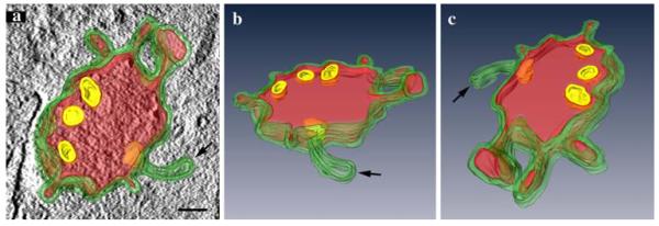
Fig. 6.
Three-dimensional reconstruction of a HDL-positive MVB. HDL-positive MVBs after 3 h of HDL internalization were analyzed by electron tomography. The 3D reconstruction of a 200-nm section demonstrates differently shaped domains of the MVB’s limiting membrane, including elongated and globular protrusions and a tubular appendix (a–c). Reaction products are colored in red. The three perspectives of the model, in a shown together with a tomographic slice, give insight into HDL-positive and negative (arrow) appendices; the intraluminal vesicles (yellow; selected ILVs were chosen for reconstruction) are devoid of reaction products (a–c). Primary magnification: 60,000×. Bar 100 nm

