Fig. 7.
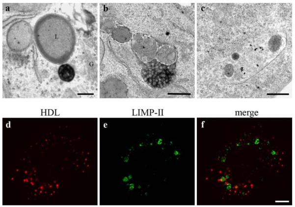
HDL is rarely found in lysosomes. Three hours after uptake of HDL-Alexa568 and DAB-photooxidation, a HDL-positive MVB is localized in the Golgi region (G) close to a lysosome (L); both, the Golgi apparatus and the lysosome are devoid of reactions (a). The very rarely observed uptake of internalized HDL in a lysosome is shown with HDL-HRP in b, and with HDL-gold in c. Poor colocalization between HDL-Alexa568 and the lysosomal marker LIMP-II (d–f) confirm these findings. Primary magnifications: 28,000× (a), 40,000× (b, c), 1,000× (d–f). Bar 250 nm (a–c), 5 μm (d–f)

