Fig. 9.
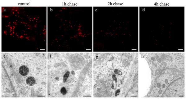
Clearance of HDL from MVBs. HepG2 cells were incubated with 50 μg/ml HDL-Alexa568 for 3 h (control) and further incubated with media containing 50 μg/ml unlabeled HDL for the indicated time points. Cells were imaged by fluorescence microscopy (a–d) or further processed for DAB photooxidation and TEM (e–h). Numbers, sizes, and staining intensities of HDL-Alexa568 positive compartments decrease time dependently (a–d). Concomitantly in the electron microscope (e–h), MVBs are reduced considerably in favor of smaller polymorphous HDL-reactive compartments (f, g). After 4-h post-incubation, nearly all MVBs are negative for HDL staining (h). Primary magnifications: 1,000× (a-–d), 70,000x (e, f), 55,000x (g), 35,000x (h). Bar 5 μm (a–d), 200 nm (e), 100 nm (f–h); N nucleus; G Golgi apparatus

