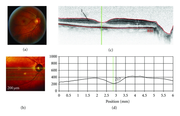
Figure 4.

SD-OCT of a patient with central retinal artery occlusion (OD). Scan parameters: infrared scan angle 30°; OCT scan angle 30°; pattern size 30°×10°, 13 sections (243 μm between B-scans). (a) The conventional fundus camera image revealed a classic “cherry red spot” and white infarctions (ischemic areas) along the major vessel arcades and around the macula. (b) SD-OCT en-face image centered approximately 2° below the fovea. The green arrow shows the position of the scan line used to generate the cross-sectional retinal OCT image (i.e., (c)). (c) The cross-sectional retinal image showed a thickening and increased reflectance (arrow “1”) of the inner retinal layers. (d) The cross-sectional retinal thickness profile revealed increased retinal thickness that was especially apparent as an exaggerated foveal pit (indicating swelling of the parafoveal retina).
