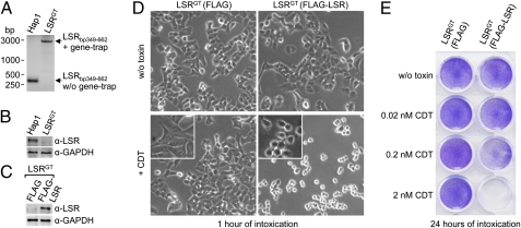
Fig. 2.
LSR is essential for intoxication of HAP1 cells with CDT. (A) PCR amplification of a region of the first intron of the LSR gene (bp 349–662) with genomic DNA isolated from HAP1 and from subcloned LSRGT cells. PCR products [with (+) and without (w/o) gene-trap insert] are shown. (B) Whole-cell lysates of HAP1 and LSRGT cells were subjected to SDS/PAGE and Western blotting, followed by detection of LSR with a specific anti-LSR antibody (α-LSR). Equal loading of proteins was verified by detection of GAPDH with a specific antibody (α-GAPDH). (C) SDS/PAGE and Western blotting with whole-cell lysates from LSRGT cells transduced with FLAG-only or FLAG-LSR retroviruses followed detection of LSR with a specific anti-LSR antibody (α-LSR). (D) FLAG- and FLAG-LSR–transduced LSRGT cells were intoxicated with 20 nM CDT or were left untreated. Cell morphology was analyzed microscopically (using the 10× objective). (E) Intoxication of FLAG- and FLAG-LSR–transduced LSRGT cells was performed with increasing concentrations of CDT (0, 0.02, 0.2, and 2 nM) as indicated, before the removal of detached cells by washing and staining of attached cells by Crystal violet dye.

