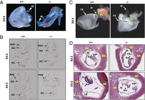
Fig. 2.
Lack of chorioallantoic attachment in FoxO1-null embryos was not due to cardiovascular malformations. (A) Normal vascular development in FoxO1-WT (+/+) and -null (−/−) YS at E9.0. Note a blood-containing honeycomb-like vascular plexus (red arrowheads) in both WT and null YS. (B) Immunohistochemical analyses for α-endomucin in WT (+/+) and FoxO1-null (−/−) embryos at the indicated developmental stages revealed normal cardiovascular development at E9.0. Note abnormal vascular (such as DA) and endocardial morphogenesis in null embryo compared with WT littermates at E9.5. Red arrowhead denotes a disrupted vessel in null mice. DA, PHV, and EC are as in Fig. 1. (C) FoxO1-WT (+/+) and -null (−/−) embryos were isolated at E8.5 and photographed without detaching from placenta (PL) and yolk sac (YS). Note that the allantois (Al) of WT, but not the null littermates is attached to the chorion (arrow). (D) H&E staining of E8.5 and E9.0-WT and -null littermates within uteri revealed lack of chorioallantoic attachment in FoxO1-null embryos. Small arrow(s) indicates chorioallantoic attachment in FoxO1-WT embryos. Al, YS, chorion (Ch), amnion (Am), uterine decidua (UD), and blood island (red box, magnified in Fig. S3) are indicated.

