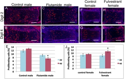
Fig. 4.
AR and ER have digit-specific effects on cell proliferation. (A–D and F–I) BrdU immunolocalization (red) and DAPI staining (blue) of longitudinal sections through proximal and middle phalanges of 2D and 4D of right hindlimbs at E16. Phalanges are oriented with proximal to the left. (Scale bars: 50 μm.) (E and J) Mitotic indices calculated from sections as represented in A–D and F–I (n = 4 embryos per group) show that antiandrogen treatment (flutamide) decreases cell proliferation in 4D of males (E), whereas antiestrogen treatment (fulvestrant) increases cell proliferation in 4D of females (J). Error bars show ± SEM. *P < 0.05.

