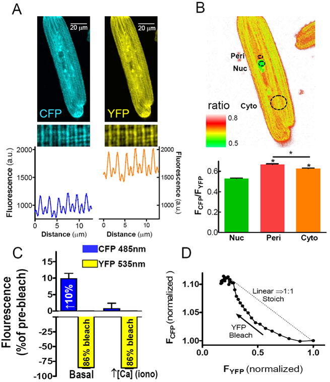
Figure 3.

Localization of Camui in cardiomyocytes. (A) Fluorescent confocal microscopy of CFP and YFP emission signals from myocytes expressing Camui. A sample line scan confirms Camui association with myofilaments. (B) A sample ratio plot shows localized differences in Camui activation in the nucleus (Nuc), perinuclear space (Peri) and cytoplasm (Cyto) at baseline conditions. (C) Selective photobleach of the YFP acceptor results in increased donor fluorescence, which is ablated by activation of CaMKII via increased [Ca2+]I. (D) CFP fluorescence is not enhanced by YFP photobleach in a 1:1 stoichiometric manner.
