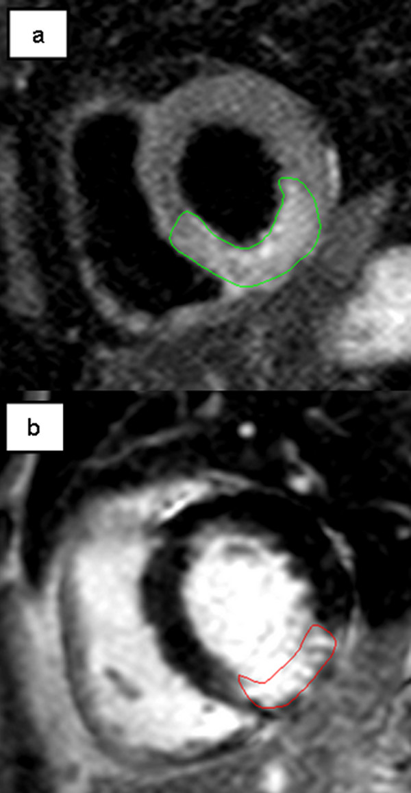
Figure 2.

Patient with inferior/inferolateral STEMI due to subtotal stenosis of right coronary artery. a. T2-weighted image acquisition for the detection of myocardial edema (green contours), corresponding to the area at risk. b. Late enhancement CMR imaging showing infarcted area (red contours). Myocardial salvage can be calculated by comparing the area at risk in T2-weighted and infarct size in late enhancement images.
