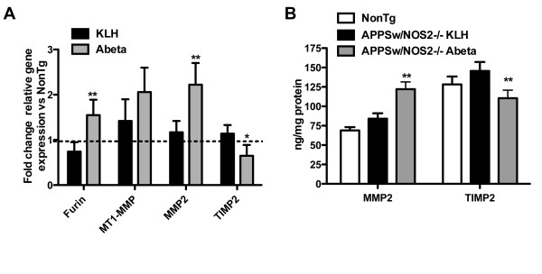
Figure 2.
Active Aβ vaccination in APPSw/NOS2-/- mice activates the MMP2 system. Panel A shows the average #177; SEM) fold change in mRNA for components of the MMP2 system in APPSw/NOS2-/- mice vaccinated with either KLH or Aβ for 4 months. Furin and MMP2 are all significantly increased following vaccination while TIMP2 is significantly decreased. The dashed line indicates the average mRNA of the non-transgenic control mice. * indicates P < 0.05, ** indicates P < 0.01 compared to KLH vaccinated APPSw/NOS2-/- mice. Panel B shows the average #177; SEM) protein levels of MMP2 and TIMP2 as measured by sandwich ELISA. Data are normalized to the protein concentration of the tissue homogenate. ** indicates P < 0.01 compared to KLH vaccinated APPSw/NOS2-/- mice.

