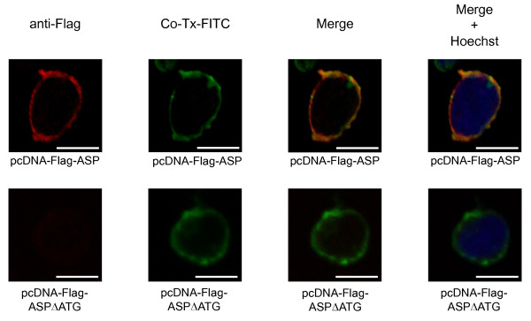
Figure 3.
HIV-1 ASP localizes to the membrane. Jurkat cells were transfected with pcDNA-Flag-ASP expressing ASP tagged with the Flag epitope to its N-terminal end or with pcDNA-Flag-ASPΔATG. Localization of ASP to the membrane was visualized by confocal microscopy using FITC-Co-Tx and immunostaining with a primary anti-Flag antibody, followed by a secondary antibody coupled to Alexa Fluor 568. Nuclei were labelled with Hoechst. White bars correspond to a scale of 10 μm.

