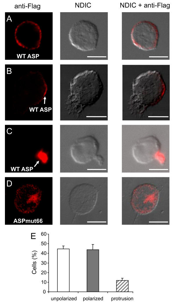
Figure 4.
Cellular localization of WT ASP and ASP-mut66 in transfected Jurkat T cells. Jurkat cells transfected with pcDNA-Flag-ASP (A-C) or pcDNA-Flag-ASPmut66 (D) were layered on glass slides, fixed, permeabilized, and stained with fluorescence-labelled antibodies as described in Figure. 3. The morphology of the cell was assessed by Normaski differential interference contrast (NDIC). White bars correspond to a scale of 10 μm. (E) Percentage of the total transfected cells with ASP showing an unpolarized distribution (white bar), a polarized location (grey bar), or a localization into membrane protrusion (hashed bar). A total of 206 cells from three separate experiments were scored.

