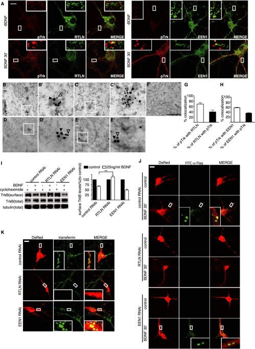
FIGURE 4:
Retrolinkin is required for BDNF-triggered TrkB internalization. (A) DIV10 hippocampal neurons were starved for 2 h and incubated with 25 ng/ml BDNF for 30 min, fixed, and immunostained with antibodies to pTrk and retrolinkin or endophilin A1. Scale bar, 10 μm. (B, C) Representative examples of double immunogold labeling of adult mouse hippocampus with antibodies to pTrk (18 nm) and retrolinkin (12 nm). (B′, C′) Higher magnification of boxed areas in B and C, respectively. (D, E) Representative examples of double immunoEM labeling of pTrk (18 nm) and endophilin A1 (12 nm). (D′, E′) Higher magnification of boxed areas in D and E, respectively. (F) Negative control with secondary antibodies only. Scale bar, 350 nm in B–F, 100 nm in B′–E′. (G) Quantification of the colocalization between pTrk and retrolinkin in immunoEM. Data represent means ± SEM (n = 3). (H) Quantification of the colocalization between pTrk and endophilin A1 in immunoEM. Data represent means ± SEM (n = 3). (I) Cortical neurons infected with Lentivirus expressing shRNA were treated with BDNF (25 ng/ml) for 30 min. TrkB internalization was then measured by cleavable surface biotinylation. Surface TrkB was detected by Western blotting with anti-panTrk. Total TrkB and tubulin were detected by Western blotting of whole-cell lysates. The quantified immunoblot bands are shown (right). Values were normalized to the respective TrkB levels of control (**p < 0.01, n = 3). (J) Hippocampal neurons transfected with FLAG-TrkB and shRNA expression constructs were treated with BDNF (25 ng/ml) for 30 min. Internalized TrkB was labeled with FITC–α-FLAG and analyzed by confocal microscopy. (K) Hippocampal neurons transfected with shRNA constructs for 3–4 d were starved for 2 h, followed by incubation with 10 μg/ml Alexa Fluor 488–conjugated transferrin for 1 h. Neurons were fixed and analyzed by confocal microscopy. EEN1, endophilin A1; RTLN, retrolinkin.

