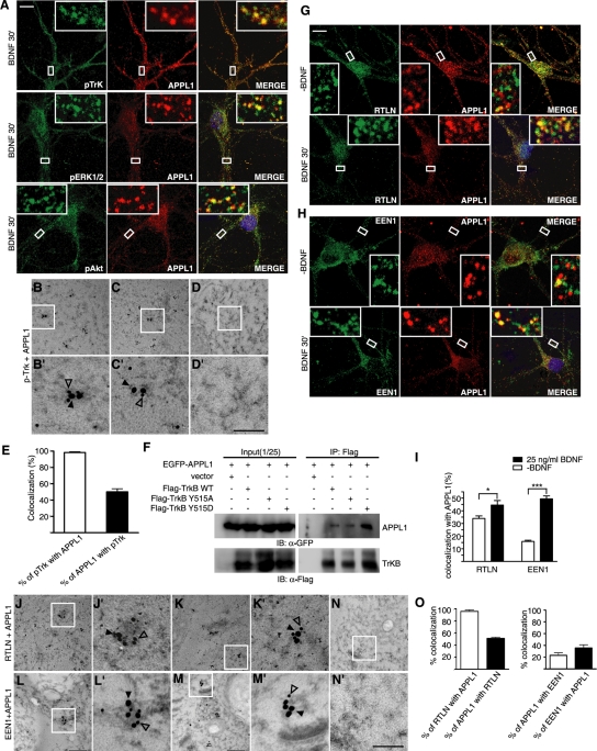
FIGURE 5:
Activated TrkB traffic through APPL1 endosomes. (A) Localization of pTrk, pERK1/2, and pAkt to APPL1-positive endosomes. DIV10 hippocampal neurons were starved for 2 h, stimulated with BDNF (25 ng/ml) for 30 min, and double stained with different antibodies. Scale bar, 10 μm. (B, C) Representative examples of double immunogold labeling of adult mouse hippocampus with antibodies to pTrk (18 nm) and APPL1 (12 nm). (D) Negative control with secondary antibodies only. (B′–D′) Higher magnification of boxed areas in B–D, respectively. Scale bar, 350 nm in B–D, 100 nm in B′–D′. (E) Quantification of the colocalization of pTrk with APPL1 in immunoEM. Data represent means ± SEM (n = 3–5). (F) CoIP of lysates of HEK293 cells overexpressing FLAG-tagged TrkB and EGFP-APPL1 with immobilized FLAG antibody. Immunoblot was probed with antibodies to FLAG and GFP. (G, H) The colocalization of retrolinkin or endophilin A1 with APPL1 was increased upon BDNF treatment. DIV10 hippocampal neurons were starved for 2 h, stimulated with BDNF (25 ng/ml) for 30 min, and double stained with antibodies to APPL1 and retrolinkin or endophilin A1. Scale bar, 10 μm. (I) Quantification of the colocalization between APPL1 and retrolinkin or endophilin A1. Data represent means ± SEM (n = 12–20) (*p < 0.05; ***p < 0.001). (J, K) Representative examples of double immunogold labeling of adult mouse hippocampus with antibodies to retrolinkin (18 nm) and APPL1 (12 nm). (L, M) Representative examples of double immunogold labeling with antibodies to endophilin A1 (12 nm) and APPL1 (18 nm). (N) Negative control with secondary antibodies only. (J′–N′) Higher magnification of boxed area in J–N, respectively. Scale bar, 350 nm in J–N, 100 nm in J′–N′. (O) Quantification of the colocalization of retrolinkin or endophilin A1 with APPL1 in immunoEM. Data represent means ± SEM (n = 3–5). EEN1, endophilin A1; RTLN, retrolinkin.

