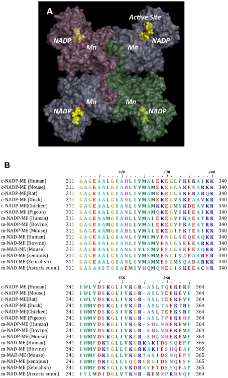
Figure 1. Nucleotide-binding site of c-NADP-ME.
(A) Tetramer of pigeon c-NADP-ME (PDB code 1GQ2). The active site with Mn2+ and NADP+ in each subunit is indicated. NADP+ in the active site is colored yellow, and the Mn2+ ion is red; they are displayed in the sphere model. (B) Multiple sequence alignments of three clusters of malic enzyme isoforms around the nucleotide-binding region of the active site. Amino acid sequences of malic enzymes were searched using BLAST [34], and the alignments were generated by Clustal W [35]. This figure was generated using the BioEdit sequence alignment editor program [36]. (C) The binding mode of NADP+ in the active site of c-NADP-ME (PDB code 1GQ2). Ser346, Lys347 and Lys362 are colored green, purple and pink, respectively, and they are shown in a ball-and-stick model. The yellow dashed lines represent the polar contacts between amino acid residues and NADP+. These figures were generated using PyMOL (DeLano Scientific LLC, San Carlos, CA).

