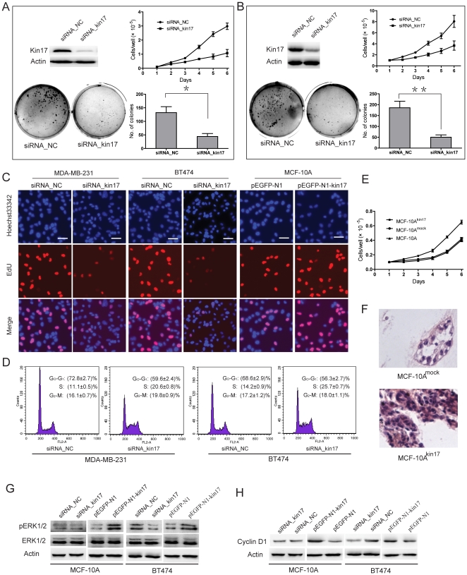
Figure 2. Association of kin17 expression with cell proliferation, colony formation, DNA replication and cell cycle progression.
MDA-MB-231 (A) and BT474 cells (B) transfected with siRNA_kin17 showed reduced growth rate and colony formation compared to the controls. * p<0.05, ** p<0.01. (C) Knockdown of kin17 expression inhibited DNA replication in MDA-MB-231 and BT474 cells compared to controls as determined by the EdU incorporation assay. Elevated expression of kin17 increased DNA replication in MCF-10A cells. (D) Cell cycle phase distributions were analyzed in a FACScalibur flowcytometer. These experiments were repeated 3 times, and the symbols represent the mean values of triplicate tests (mean ± SD). (E) Overexpression of kin17 following transfection with pEGFP-N1-kin17 promoted cell proliferation in MCF-10A cells. Cells positive for the EGFP-N1-kin17 were designated MCF-10Akin17, while cells transfected with pEGFP-N1 vector were designated MCF-10Amock. (F) Overexpression of kin17 disrupted acinar organization and blocked luminal clearance in MCF-10A cells. Western blotting analysis of autophosphorylation of ERK1/2 (G) and cyclin D1 expression (H). The experiment was independently repeated at least 3 times.

