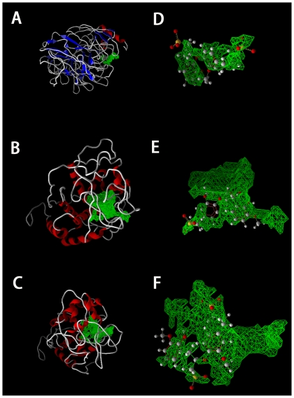
Figure 1. Binding pockets and binding orientations of lignin in the best docking Lac-lignin, LiP-lignin and MnP-lignin complexes.
Panels A, B and C display the binding pockets of Lac, LiP and MnP, respectively, whereas panels D, E and F show the binding orientations of Lac, LiP and MnP, respectively. The 3D structures of Lac, LiP and MnP are represented in Cartoon style. The green grids show the binding pockets of lignin-enzymes. The lignin is clearly showed in ball and stick model (colored by element: gray, carbon; red, oxygen; white, hydrogen; yellow, sulfur).

