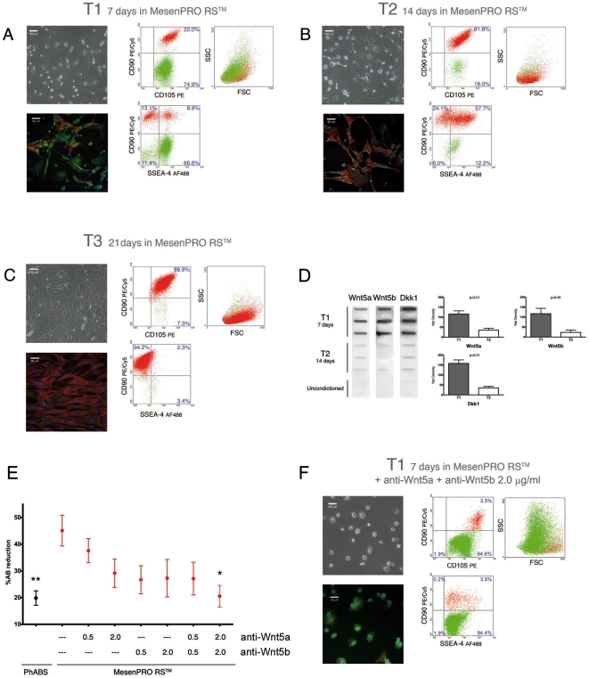
Figure 3. Mesenchymal differentiation of MPCs and Wnt signalling.
(A) After 7 days of culture (T1) in differentiating medium some spindle-shaped cells were detectable (Leica DM-RB, HBO-50W Fluorescence 400X, merging by Leica CW4000 software). SSEA-4 (green) and CD90 (red, blue stain for DAPI) immunofluorescence and flow cytometry allowed the identification of a portion of MSCs as early MSCs (SSEA-4 and CD90 positive). (B) After a further 7 days (T2) cultures showed a reduced population of MPCs alongside an increased population of early MSCs. (C) Confluence of spindle-shaped cells was obtained after 3 weeks of differentiation (T3). Immunofluorescence showed that cells were completely negative for SSEA-4 while expressing CD90. Flow cytometry confirmed the unique phenotype of late MSCs (SSEA-4negCD105brightCD90bright). (D) Slot-Blot of conditioned media revealed intense secretion of Wnt5a and Wnt5b at T1 and very low secretion at T2. Similarly, Dkk1 was highly secreted at T1 and reduced at T2. (E) Autocrine/paracrine actions of Wnt5a and Wnt5b were redundant and differentiation was significantly inhibited with high doses (expressed in µg/ml) of specific antibodies (* p<0.05, ** p<0.01). (F) Immunoblocking of Wnt5a and Wnt5b activity, during differentiation, resulted in the retention of MPC phenotype in about 95% of the cells, after 7 days of induction.

