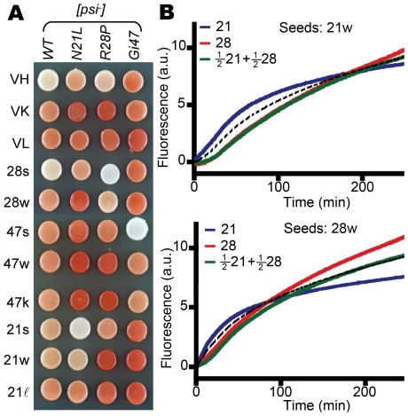
Figure 4. Distinct structures of [PSI] strains.
(A) Yeast harboring a [PSI] strain (labeled on the left) is mated with different genetic backgrounds (labeled on top). Homozygotes always exhibit stronger suppression of the ade2-1 nonsense mutation, resulting in lighter colony color. (B) Nucleated growth of prion fibers monitored by ThT fluorescence in vitro. Top panel: 21w seeds. Bottom: 28w seeds. 21 = 1 µM solution of Sup(1-253)(N21L) (blue); 28 = 1 µM solution of Sup(1-253)(R28P) (red). The 0.5∶0.5 mixture (green) exhibits lower ThT fluorescence at early time points compared with the averaged signal (dotted black line) of the two pure solutions.

