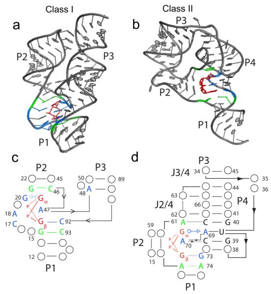
Figure 1.
Crystal structures of the class I and class II riboswitch aptamer domains bound to c-di-GMP. c-di-GMP is colored in red, nucleotides in direct contact with the ligand are show in blue, and nucleotides that stack directly above and below the ligand are shown in green. (a) Structure of the class I Vc2 aptamer from V. cholera bound to c-di-GMP (PDB ID 3MXH). (b) Structure of the class II Cac-1-2 aptamer from C. acetobutylicum bound to c-di-GMP (PDB ID 3Q3Z). (c) Binding pocket of the class I aptamer. (d) Binding pocket of the class II aptamer. Nucleotides that form base triples with residues directly contacting c-di-GMP are shown.

