
Figure 1.
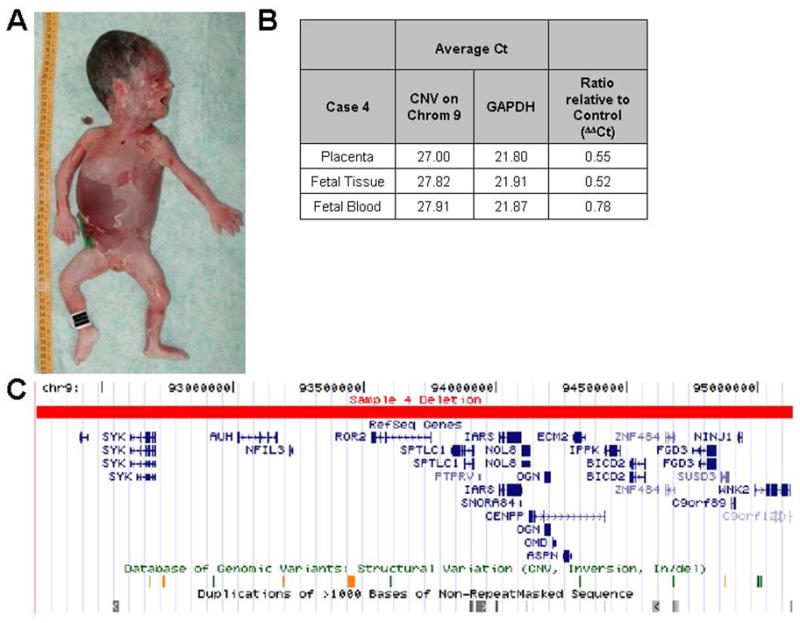
A 2.89 Mb deletion identified in case 4. A) Autopsy photograph of fetus, with complete anatomic pathology revealing no evidence of gross nor histologic abnormalities. B) SYBR Green qPCR results showing a ratio relative to control (ΔΔCt) approximating 0.5 confirming the deletion. A ΔΔCt in fetal blood was 0.78, possibly due to maternal blood contamination. C) UCSC Genome Browser view of the genomic region deleted. The deletion affected 25 known (RefSeq) genes (with complete listing as noted in Table VI). Known structural variations and segmental duplications in this region are also shown.