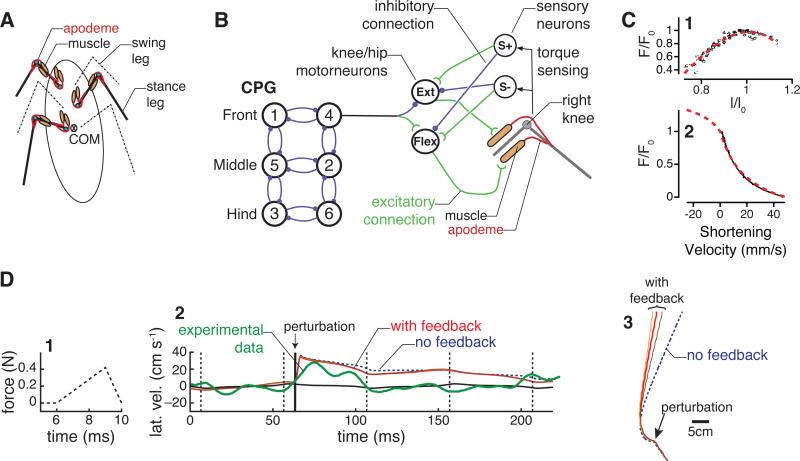
Figure 3.
Instantiation of the system of Fig. 1 in a model of insect locomotion. A Mechanical model. Extensor and flexor muscles actuate simplified “hip-knee” geometry modeling coxa-femur and femur-tibia joints. B Six hemisegments constitute a CPG oscillator network that drives motor neurons (MNs) in a feedforward manner. Joint torques monitored by campaniform sensilla modulate relative phases of MN bursts (via S+ and S- neurons), but primary environmental feedback comes from mechanical reaction forces and stretch and stretch-rate force dependence in muscles. Filled circles and open arcs respectively denote excitatory and inhibitory connections. C Forces produced by muscle depend on length (panel 1) and shortening velocity (panel 2). Data from Ahn and Full [53] shown in black; fits shown with red dashed lines. D Response of the model as diagrammed in panels A-C to a rapid lateral perturbation. 1 Time course of perturbation force. 2 Lateral velocity after the perturbation. Solid black line shows the unperturbed model. Dashed blue line shows the model with no sensory feedback, while solid orange, red, and brown lines show differing sensory feedback gains. For comparison, experimental data from [52] is overlaid with a thick green line. 3 Trajectory of the model's mass center in the horizontal plane. Feedback reduces heading change after the perturbation. Modified from [54, 55].

