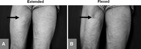
Fig. 1A–B.
The photographs show a posterior view of the thighs with the patient in the prone position. (A) When the patient was examined in the prone or standing position with the knee extended, the left hamstrings had a normal appearance compared to the opposite side. For orientation, the arrow corresponds to the area of the defect in the next image. (B) When the patient was examined in the prone or standing position with the knee flexed, the defect was clearly visible (arrow), but only with active, not passive, flexion.

