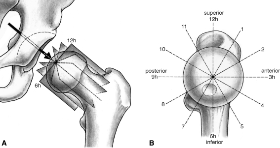
Fig. 1A–B.
(A) The radial MRI planes, which are perpendicular to the femoral head-neck axis, are defined on a sagittal oblique localizer. (B) The radial cuts rotate clockwise in 30°-intervals around the femoral head-neck axis. The alpha angle measurements were performed throughout the cranial hemisphere from 9 o’clock to 3 o’clock.

