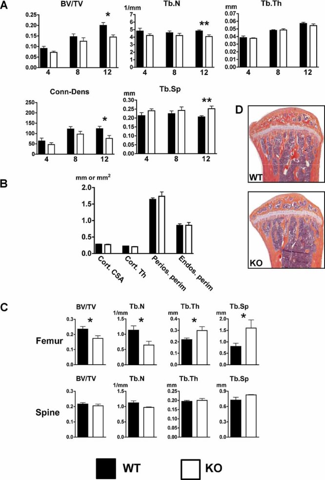
Fig. 3.

Reduced tibia and femur trabecular bone volume in 12-week-old female KO mice. (A) The tibias of 4-, 8-, and 12-week-old female WT and KO mice were analyzed by µCT. Trabecular bone volume (BV/TV), trabecular number (Tb.N), trabecular thickness (Tb.Th), connectivity density (Conn.D), and trabecular separation (Tb.Sp) were determined. (B) Tibia cortical bone analysis of 12-week-old female mice revealed that osteoblast-targeted ETAR KO did not alter cortical parameters by µCT. Cortical cross-sectional area (Cort.CSA), cortical thickness (Cort.Th), periosteal perimeter (Perios.perim), and endosteal perimeter (Endos.perim) were determined. (C) Static bone histomorphometric analysis demonstrated reduced femoral, but not spine, trabecular bone volume in 12-week-old KO female mice. (D) Representative tibias from WT and KO mice stained with H&E/orange G (*p ≤ 0.05; **p ≤ 0.01).
