Fig. 7.

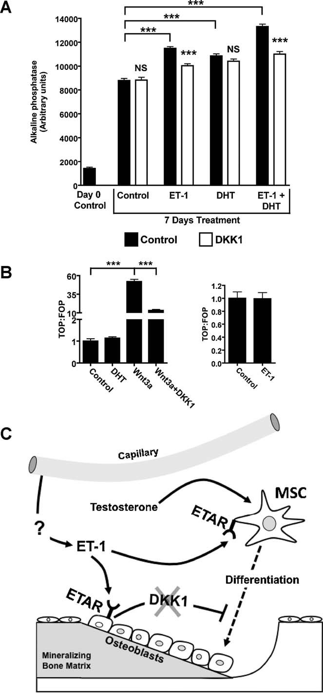
Mechanisms of ET-1 and DHT action in the osteoblast. (A) In MC3T3 osteoprogenitor cells, ET-1 (100 nM), DHT (10 nM), and combined treatment increased AP expression after 7 days of treatment. DKK1 (50 ng/mL) diminished AP staining in ET-1 and in combined ET-1 + DHT–treated cells. DKK1 did not change AP staining in DHT-treated cells. (B) Neither DHT nor ET-1 increased canonical Wnt signaling, as measured using Wnt reported vectors in isolated MC3T3 cells. Wnt3a (50 ng/mL) increased canonical Wnt signaling that was abrogated by DKK1 (50 ng/mL). (C) Model of ET-1, DHT, and DKK1 in regulating osteoblast activity. The source of ET-1 in the bone microenvironment is most likely vascular endothelial cells of adjacent capillaries. ET-1 promotes osteoprogenitor differentiation through direct mechanisms independent of Wnt activation and indirectly by lowering the tonic inhibition of DKK1 secreted in mature osteoblasts. Testosterone complements the actions of ET-1 by increasing osteoprogenitor differentiation through unclear mechanisms (*p ≤ 0.05; ***p ≤ 0.001).
