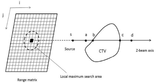
Fig. 1.

A schematic illustration of the method used to calculate the range matrix and relevant margin of a ray. Radiological path length is calculated per ray, and a kernel is applied to replace the radiological path length of a given ray with the local maximum within a distance (the lateral setup error and organ motion) of the range matrix. 3.5% of the assigned path length is used to convert to physical depth to form a margin (distal , proximal ).
