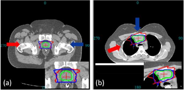
Fig. 5.

The CTV (green contour) is used to derive two bsPTVs (red and blue contours) under same specification (setup and range error) at different angles. (a) For prostate site, both bsPTV shows characteristic horn like distal shape to account for the misalignment of highly dense femur and femoral head. (b) For thoracic site, the two bsPTVs are significantly different in its shape and volume due to the difference in tissue density along their beam paths.
