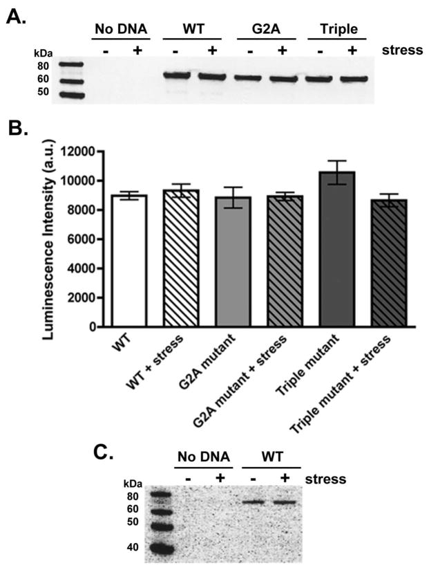
Figure 4.
PAFAH-II expression levels. (A) Representative GFP Western blot of HEK293 lysates expressing PAFAH-II constructs before and after treatment with 80 μM cumene hydroperoxide as an oxidative stressor. GFP antibodies are suitable for detection of both YFP and CFP proteins. (B) PAFAH-II construct expression in HEK293 cell lysates before and after stress, measured by relative luminescence intensity and subsequent densitometry analysis from four independent GFP Western blots. (C) Using a PAFAH-II antibody, HEK293 cells were shown to not have any detectable levels of endogenous PAFAH-II before or after stress, as seen in the “No DNA” sample (expected MW: 44 kDa). Cells expressing WT-PAFAH-II-YFP construct were used as a positive control.

