
Figure 1.
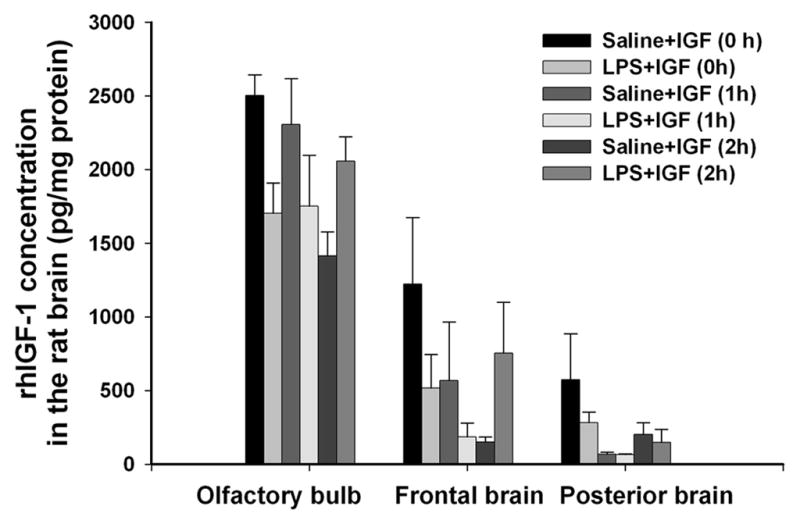
Concentrations of rhIGF-1 in different parts of the rat brain 30 min after intranasal administration of rhIGF-1 in the initial study. Intranasal infusion of rhIGF-1 was performed at 0, 1 or 2 hr after the intraceberal injection of LPS or sterile saline in P5 rats. Each group contained 5 animals. rhIGF-1 was detected in the brain of rhIGF-1-infused animals, but not in that of the vehicle (0.1% BSA)-infused animals, from either the saline- or the LPS-injected group. rhIGF-1 concentrations in the FB and the PB of saline- or LPS-injected rats brain were lower than that in the OB. No significant differences in rhIGF-1 concentrations were observed among the treatment groups within respective brain region.