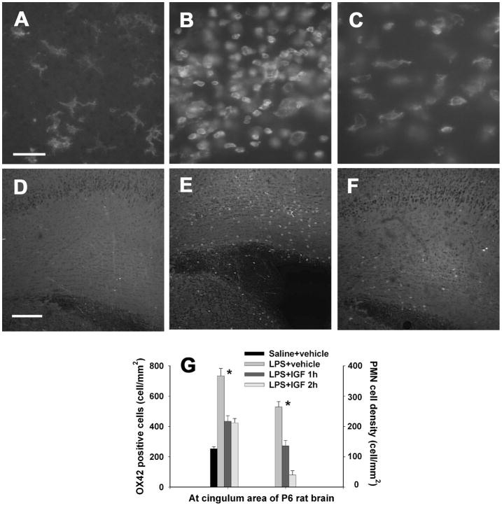
Figure 5.
Density of OX42 positive microglia (A–C) and infiltrated CD43 positive PMN cells (D–F) in the P6 rat brain. Perinatal LPS exposure resulted in a significant increase in the number of microglia, which have a round shape (B) as compared to the control rat brain (A) 24 h after the LPS exposure. IGF-1 administration at 2 hr after LPS injection reduced microglial activation (C). Most microglia in the control rat brain and some microglia in the LPS+IGF rat brain have a ramified shape. No CD43+ PMN cells were detectable in the control rat brain (D). Perinatal LPS exposure significantly increased the number of PMN cells at the cingulum area of the rat brain 24 h after LPS injection (E). IGF-1 treatment at 2 hr after LPS injection reduced PMN infiltration (F). The quantitative data of OX42+ microglia density and CD43+ PMN cell density in the rat brain are presented in G (n=6 for each grtoup). *p<0.05 vs the other groups. Scale bar: A–C, 50 μm; D–F, 200 μm.

