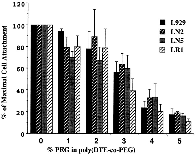
Figure 2.
Quantitative analysis of single cell attachment on poly(DTE-co-PEG carbonate) substrata shows that in all L cell types, rate of attachment markedly declined with increasing PEG content. Fluorescence-labeled (calcein-AM dye) single cell suspensions of low, intermediate, and high cohesivity were allowed to attach for 24 h on polymeric substrata containing 0–5% PEG. Rate of cell attachment was measured as fluorescence intensity at each time point. Data points represent averages of measurements made in duplicate and repeated at least three times.

