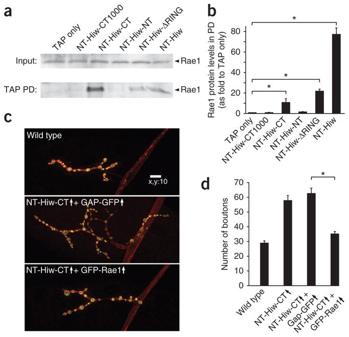
Figure 6.
Rae1 interacts with a fragment in the Hiw C-terminal region, and coexpression of Rae1 with NT-Hiw-CT suppresses the NT-Hiw-CT–induced dominant-negative overgrowth phenotype. (a) Indicated NTAP-tagged hiw transgenes (described in Fig. 5) and a TAP only transgene were expressed in neurons under the control of the BG380-Gal4 driver. Larval brain lysates from each sample were subject to IgG pulldown. Both the inputs and the IgG pulldown complexes were analyzed by western blot using antibody to Rae1. Rae1 was present in all of the pulldown complexes except for those from TAP only, NT-Hiw-CT1000 and NT-Hiw-NT. Full-length blots are presented in Supplementary Figure 10. (b) Quantification of the interaction between various Hiw transgenic proteins and Rae1. Rae1 intensities in TAP pulldown blots were normalized to both the intensities and molecular weights of the corresponding TAP pulldown proteins. (c) Representative confocal images of muscle 4 synapses stained for both DVGLUT (green) and FasII (red) in wild-type (BG380/Y;+/+; +/+), NT-Hiw-CT and GAP-GFP (BG380/Y; UAS-GAP-GFP/+; UAS-NT-hiw-CT/+), and NT-Hiw-CT and Rae1 (BG380/Y; +/+; UAS-NT-hiw-CT/UAS-GFP-Rae1) larvae. Scale bar represents 10 μm. (d) Quantification of number of boutons at muscle 4 NMJs in wild-type, NT-Hiw-CT overexpression (BG380/Y; +/+; UAS-NT-hiw-CT/+), NT-Hiw-CT and GAP-GFP, and NT-Hiw-CT and Rae1 (n = 24, 27, 32 and 26, respectively) larvae. Error bars denote s.e.m.; *P < 0.001.

