Figure 7.
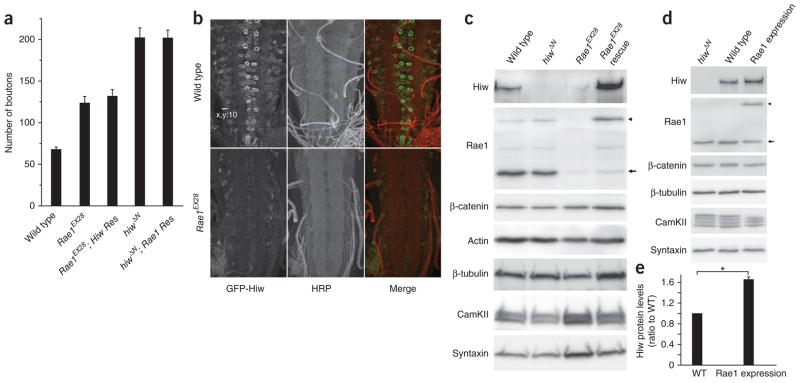
Rae1 promotes Hiw abundance. (a) Neuronal expression of neither Hiw nor Rae1 rescues the synaptic terminal overgrowth phenotype caused by the loss of function of the other gene. Quantification of the number of boutons in wild-type, Rae1EX28, Rae1EX28; Hiw rescue (Res) (Rae1EX28; elav-Gal4/UAS-GFP-hiw), hiwΔN and hiwΔN; Rae1 rescue (hiwΔN; elav-Gal4/UAS-NTAP-Rae1) larval NMJs (segment A3 muscle 6/7; n = 12, 12, 12, 7 and 11, respectively). (b) Representative confocal images of wild-type (BG380-Gal4/+; +/+; UAS-GFP-hiw/+) or Rae1 mutant (BG380-Gal4/+; Rae1EX28; UAS-GFP-hiw/+) ventral ganglions with UAS-GFP-hiw trangene expressed in neurons. The larvae were stained with antibodies to horseradish peroxidase (HRP, red) and GFP (green). Scale bar represents 10 μm. (c) Western blots of total proteins extracted from wild-type, hiwΔN, Rae1EX28 and Rae1EX28 rescue (Rae1EX28; elav-Gal4/UAS-NTAP-Rae1) larval brains probed with indicated antibodies. Full-length blots are presented in Supplementary Figure 11. (d) Western blot analysis on total proteins extracted from hiwΔN, wild-type (C155-Gal4/+) and Rae1 expression (C155-Gal4/+; UAS-NTAP-Rae1/+) larval brains. Full-length blots are presented in Supplementary Figure 12. (e) Quantification of Hiw protein levels in the wild-type and the Rae1 expression larval brains (*P < 0.001, based on seven independent experiments). Arrows indicate a 38-kDa endogenous Rae1 protein and the arrowheads indicate the 58-kDa transgenic NTAP-Rae1 protein. Error bars denote s.e.m.

