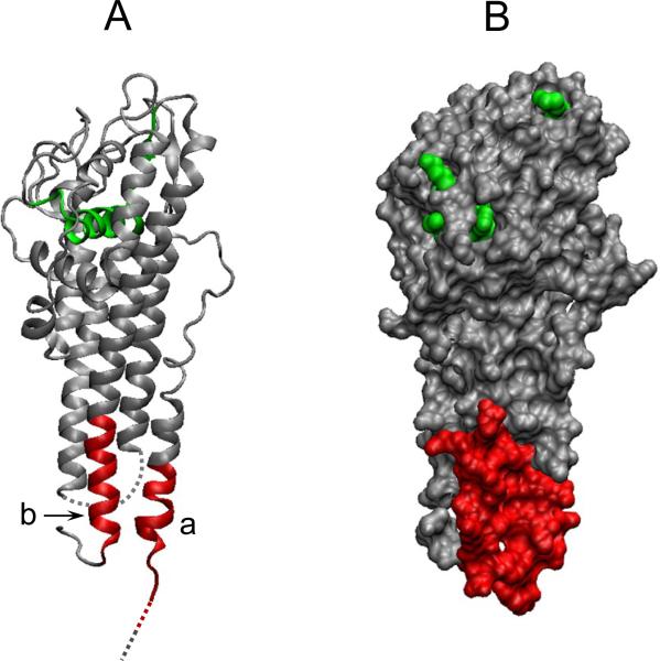
Figure 4. Spatial position of epitopes of VlsE for which a differential antibody response is found in the PLDS patient group.
A ribbon diagram (A) and an orthographic molecular surface representation (B) of VlsE monomer are depicted using the VMD molecular graphics program, based on NCBI's 3D-structure database coordinates. Two specific epitopes of the protein believed to be differentially targeted by antibodies in PLDS patients are shown in red: a, VlsE21-31: SQVADKDDPTNKFYQSVIQLGNGF. b, VlsE336: LRKVGDSVKAASKE. Parts of the protein that were missing from the 3D-structure database coordinates are represented as dashed lines in the ribbon diagram. The sequence representing the IR6 epitope of VlsE (used in C6 ELISA) is shown in green. The bottom of the figure represents the membrane proximal region.

